The University of Toledo
The University of Toledo Digital Repository
Theses and Dissertations
2013
The regulatory role of mixed lineage kinase 4 beta
in MAPK signaling and ovarian cancer cell invasion
Widian F. Abi Saab
The University of Toledo
Follow this and additional works at: http://utdr.utoledo.edu/theses-dissertations
Recommended Citation
Abi Saab, Widian F., "The regulatory role of mixed lineage kinase 4 beta in MAPK signaling and ovarian cancer cell invasion" (2013).
Theses and Dissertations. Paper 2.
This Dissertation is brought to you for free and open access by The University of Toledo Digital Repository. It has been accepted for inclusion in Theses
and Dissertations by an authorized administrator of The University of Toledo Digital Repository. For more information, please see the repository's
About page.
A Dissertation
entitled
The Regulatory Role of Mixed Lineage Kinase 4 Beta in MAPK Signaling and Ovarian
Cancer Cell Invasion
by
Widian F. Abi Saab
Submitted to the Graduate Faculty as partial fulfillment of the requirements for the
Doctor of Philosophy Degree in Biology
_________________________________________
Dr. Deborah Chadee, Committee Chair
_________________________________________
Dr. Douglas Leaman, Committee Member
_________________________________________
Dr. Fan Dong, Committee Member
_________________________________________
Dr. John Bellizzi, Committee Member
_________________________________________
Dr. Max Funk, Committee Member
_________________________________________
Dr. Robert Steven, Committee Member
_________________________________________
Dr. William Taylor, Committee Member
_________________________________________
Dr. Patricia R. Komuniecki, Dean
College of Graduate Studies
The University of Toledo
May 2013
Copyright 2013, Widian Fouad Abi Saab
This document is copyrighted material. Under copyright law, no parts of this document
may be reproduced without the expressed permission of the author.
An Abstract of
The Regulatory Role of Mixed Lineage Kinase 4 Beta in MAPK Signaling and Ovarian
Cancer Cell Invasion
by
Widian F. Abi Saab
Submitted to the Graduate Faculty as partial fulfillment of the requirements for the
Doctor of Philosophy Degree in Biology
The University of Toledo
May 2013
Mixed lineage kinase 4 (MLK4) is a member of the MLK family of mitogenactivated protein kinase kinase kinases (MAP3Ks). As components of a three-tiered
signaling cascade, MAP3Ks promote activation of mitogen-activated protein kinase
(MAPK), which in turn regulates different cellular processes including proliferation and
invasion. Here, we show that the beta form of MLK4 (MLK4β), unlike its close relative,
MLK3, and other known MAP3Ks, negatively regulates the activities of the MAPKs,
p38, ERK and JNK, even in response to stimuli such as sorbitol or TNFα. MLK4β also
negatively regulates basal, but not TNFα-induced, NF-κB activity. Moreover, MLK4β
undergoes autophosphorylation and has kinase activity towards histone H2A, but has no
kinase activity towards the MAP2K, MEK4/SEK1, a known substrate for MLK3 and
other MAP3Ks. Furthermore, MLK4β interacts with MLK3 and inhibits MLK3
activation. In addition, MLK4 blocks matrix metalloproteinase-9 gelatinase activity and
invasion in SKOV3 ovarian cancer cells, both of which are cellular responses that require
MLK3. Collectively, our data establish MLK4β as a novel suppressor of MLK3
activation, MAPK signaling and cell invasion.
iii
This work is dedicated to my dad, Fouad Abi Saab, and mom, Nabila Abi Saab,
who sacrificed a lot to provide a good education for my brother and me. I most certainly
would not be where I am today if it wasn’t for them.
I also dedicate this work to my brother (Rawad), my grandmas (Fayza and
Samia), my aunts (Thouraya, Feryal, Noha and Sonia) and all my cousins (Yara, Ziad,
Lama, Wahid, Tamara and Faisal). However, a special dedication goes to my beloved
Grandma, Fayza Darweesh, who is my role model. She is my inspiration and the source
of my strength and has always been my number one supporter. Her words and constant
encouragement are my driving force to move forward in life.
I would also like to grab this opportunity to thank my dearest friends (Alexis, Ani,
Celia, Hadil, Hashem, Meenakshi, Mirella, Nancy and Natalya) who’ve been extremely
encouraging and supportive throughout my Ph.D. program.
Acknowledgements
First, I would like to thank my advisor, Dr. Chadee, who had given me the chance
to be here and who taught me most of what I currently know in this field. Dr. Chadee is a
very supportive and positive person and creates a very amiable environment for her
students. In addition to being successful in her field, she is also extremely compassionate
and understanding. She was very supportive especially during hard times and for that I’ll
be forever grateful. Not only is Dr. Chadee successful in her career, but she also has an
exemplary sense of humanity which makes her a great role model for me.
I would also like to thank my committee members Dr. Douglas Leaman, Dr. Fan
Dong, Dr. John Bellizzi, Dr. Max Funk, Dr. Robert Steven and Dr. William Taylor for
their constant input and guidance. I especially thank Dr. Taylor, Dr. Leaman and Dr.
Dong, for their technical support in a number of experiments.
I would like to especially thank Cathy (Dr. Yu Zhan) for teaching me most of the
techniques in lab and for being a good friend. Special thanks to Natalya Blessing for
being a wonderful lab mate and friend. I would also like to thank Meenakshi Bhansali for
her amazing friendship and support. Last but not least, I would like to thank Dr. Leah
Rider, Jenny, Alan, Peter, Sneha, April and Kyoung for being such good friends and for
adding a joyful and pleasant atmosphere to our working environment.
v
Table of Contents
Abstract .............................................................................................................................. iii
Acknowledgements ..............................................................................................................v
Table of Contents ............................................................................................................... vi
List of Figures .................................................................................................................... ix
List of Abbreviations ......................................................................................................... xi
1
Introduction………………………………………………………………………..1
1.1 The Mitogen-activated protein kinase signaling cascade ...............................1
1.2 Characteristics and functions of MAPK proteins…………………………….3
1.2.1 The ERK1/2 pathway………………………………………………..3
1.2.2 The JNK pathway…………………………………………………...7
1.2.3 The p38 pathway…………………………………………………...11
1.2.4 The ERK5 pathway………………………………………………...14
1.3 The matrix metalloproteinases……………………………………………….15
1.4 The MAP2Ks………………………………………………………………...18
1.5 The MAP3Ks………………………………………………………………...20
1.5.1 The MEKK group………………………………………………….21
1.5.2 The Raf MAP3Ks………………………………………………….23
1.5.3 The TAK1 MAP3K group…………………………………………25
1.5.4 The TAO/Tpl2 and Mos MAP3K groups………………………….27
1.5.5 The MLK family of MAP3Ks……………………………………..27
vi
1.5.5.1 The DLK subgroup ...........................................................28
1.5.5.2 The ZAK subgroup………………………………………30
1.6 The MLK subfamily…………………………………………………………31
1.6.1 MLK1 and MLK2………………………………………………….32
1.6.2 MLK3 activation…………………………………………………...33
1.7 MLK3 signaling……………………………………………………………...36
1.7.1 MLK3 signaling in cancer…………………………………………38
1.8 MLK4: characteristics and function………………………………………….39
1.9 Significance…………………………………………………………………..40
2
Materials and Methods ...........................................................................................42
2.1 Cell culture…………………………………………………………………...42
2.2 Expression vectors…………………………………………………………...43
2.3 Plasmids and siRNA transfections…………………………………………...43
2.4 Immunoblotting………………………………………………………………45
2.5 Preparation of whole cell extracts and treatments…………………………...47
2.6 Immunoprecipitation…………………………………………………………47
2.7 MLK4β kinase assay…………………………………………………………48
2.8 Cell proliferation assay………………………………………………………49
2.9 Luciferase assay……………………………………………………………...50
2.10 Invasion assay………………………………………………………………50
2.11 Gelatin zymography………………………………………………………...51
3
Results……………………………………………………………………………52
3.1 The role of MLK4β in p38 signaling………………………………………...52
vii
3.1.1 The effect of ectopic expression of MLK4β on p38 activation…...52
3.1.2 The effect of endogenous MLK4 on the activation of p38. ………54
3.2 The effect of MLK4 on MEK3/MEK6 activation….………………………..56
3.3 The role of MLK4β in NF-κB signaling……………………………………..57
3.4 Comparison of the effects of MLK4β and MLK3 on p38 activation ……….60
3.5 The effects of MLK3 and MLK4 on ERK and JNK activation……………..62
3.6 MLK4β is not an upstream activator of MEK4……………………………...65
3.7 MLK4β kinase activity……………………………...……………………….67
3.8 The effect of MLK4β on MLK3 activation………………………………….69
3.9 The correlation between MLK4β expression and active MLK3 in different cell
lines……………………………………………………………...……………….72
3.10 MLK4β associates with MLK3……………………………………………..75
3.11 The effect of MLK4 on cell proliferation…………………………………..77
3.12 MLK3 is required for cell invasion in ovarian cancer cells………………...79
3.13 MLK4β inhibits SKOV3 cell invasion……………………………………..81
3.14 MLK3 regulates MMP-2 and MMP-9 enzyme activity…………………….82
3.15 MLK4β reduces MMP-9 activity in SKOV3 cells…………………………85
4
Discussion………………………………………………………………………..86
References……………………………………………………………………………….95
viii
List of Figures
1-1
The MAPK signaling cascade ..................................................................................2
1-2
The Ras/Raf/ERK1/2 signaling pathway .................................................................6
1-3
JNK-mediated apoptosis…………………………………………………………10
1-4
The p38 MAPK signaling pathway………………………………………………13
1-5
MMP-2 and -9: structure and activation…………………………………………17
1-6
The NF-κB pathway……………………………………………………………...26
1-7
Signaling of the DLK family of MAP3Ks……………………………………….29
1-8
The structural domains of MLKs………………………………………………...31
1-9
Model mechanism of MLK3 activation by Cdc42………………………………35
3-10
MLK4β expression inhibits basal and stimulus-induced p38 activation ...............54
3-11
Elevated active p38 in MLK4 knockdown cells…………………………………55
3-12
MLK4 negatively regulates MEK3/MEK6 activation…………………………...57
3-13
MLK4β negatively regulates basal NF-κB activation but has no effect on TNFα-
induced NF-κB signaling……………………………………..………………………….59
3-14
MLK4β, unlike MLK3, inhibits activation of p38 ………………………………61
3-15
MLK3 promotes the activation of ERK and JNK in SKOV3 and HEY1B cells...62
3-16
MLK4 negatively regulates ERK and JNK activation…………………………...64
3-17
MLK4β does not phosphorylate Thr261 on GST-SEK1-KR……………………66
3-18
MLK4β: autophosphorylation and substrate specificity…………………………68
3-19
MLK4β inhibits induced MLK3 activation……………………………………...69
ix
3-20
MLK4 inhibits the basal activation of MLK3 in SKOV3 cells………………….71
3-21
Correlation between MLK4β expression and active MLK3.. …………...............74
3-22
MLK4β associates with MLK3………………………………………………......76
3-23
MLK4 has no effect on HCT116 cell proliferation……………………………...78
3-24
MLK3 is essential for SKOV3 and HEY1B cell invasion……………………….80
3-25
MLK4β reduces the invasion of SKOV3 cells..…………………………………82
3-26
MLK3 mediates MMP-2 and MMP-9 activation in SKOV3 and HEY1B cells by a
mechanism that involves ERK and JNK………………….……………………………..84
3-27
MLK4β reduces MMP-9 activity in SKOV3 cells………………………………85
4-28
Schematic diagram illustrating the role of MLK4β in MAPK signaling………..94
x
List of Abbreviations
Akt1............................Rac-alpha serine/threonine kinase
APS ............................Ammonium persulfate
AP-1 ...........................Activator protein 1
ASK............................Apoptosis signal-regulating kinase
ATF2 ..........................Activating transcription factor 2
ATM………………..Ataxia telangiectasia
ATP ...........................Adenosine triphosphate
Bax .............................Bcl2-associated X
Bcl-2 ...........................B-cell lymphoma 2
BSA ............................Bovine serum albumin
CBD ...........................Collagen binding domain
CR ..............................Conserved region
CRIB ..........................Cdc42/Rac interactive binding protein
DTT ............................Dithiothreitol
DLK ...........................Dual leucine zipper-bearing kinase
DNA ...........................Deoxyribonucleic acid
DSP ............................Dual specificity phosphatases
ECM ...........................Extracellular matrix
EMT ...........................Epithelial-mesenchymal transition
EGF ............................Epidermal growth factor
ERK ...........................Extracellular signal-regulated kinase
FADD.……….……..Fas-associated death domain protein
FasL............................Fas ligand
FBS ............................Fetal bovine serum
FFA…………………Free fatty acids
FGD1……………….FYVE, RhoGEF and PH domain-containing protein 1
GADD45 ....................Growth arrest and DNA damage-inducible 45
GDP………………...Guanosine diphosphate
GEF ............................Guanosine nucleotide exchange factor
xi
Grb2 ...........................Growth factor receptor binding protein 2
GST ............................Glutathione S-transferase
GTP ............................Guanosine triphosphate
h……………………..hours
HBx…………………Hepatitis B x antigen
HPK1..........................Hematopoietic protein kinase 1
IκBα ...........................Inhibitor of kappa B alpha
IKK ............................IκB kinase
IKKK..........................IκB kinase kinase
IP ................................Immunoprecipitation
JIP1 ............................JNK-interacting protein 1
JNK ...........................c-Jun N-terminal kinase
LPS .............................Lipopolysaccharide
LZ ...............................Leucine zipper
KSR ............................Kinase suppressor of ras
MAPK ........................Mitogen activated protein kinase
MAPKAP-K...............MAPK-activated protein kinase
MAP2K ......................MAPK kinase
MAP3K ......................MAPK kinase kinase
MEK...........................MAPK/ERK kinase
MEKK ........................MEK kinase
Met .............................Methionine
MLK ...........................Mixed lineage kinase
MLTKα ......................Mitogen-activated protein triple kinase alpha
MKP ...........................MAPK phosphatase
MMP ..........................Matrix metalloproteinase
MNK ..........................MAPK interacting kinase
MP-1 ..........................MEK partner 1
MSK ...........................Mitogen and stress activated kinase
ND………………….Neuro-D
NF ..............................Neurofibromatosis
NF-κB ........................Nuclear factor kappa-light-chain enhancer of activated B cells
NGF............................Neuron growth factor
PAK1..........................p21-GTPase activated kinase 1
PARP……………….poly (ADP-ribose) polymerase (PARP)
PB1.............................Phox/Bem1P
PBS ............................Phosphate buffered saline
PHD………………...Plextrin-homology domain
xii
PMSF .........................Phenylmethylsulphonyl fluoride
PP2A ..........................Serine/threonine protein phosphatase 2A
PP5 .............................Protein phosphatase 5
Pro ..............................Proline
PTP............................. protein tyrosine phosphatase
PVDF .........................Immobilon-P Polyvinylidene Flouride
ROS………………...Reactive oxygen species
RSK ............................p90 ribosomal S6 kinase
RTK............................Receptor tyrosine kinase
SAP1………………..Sodium-associated protein 1
SAM ...........................Sterile-alpha-motif
SAPK .........................Stress-activated protein kinase
SCG………………...Superior cervical ganglion
Ser ..............................Serine
SH ..............................Src homology
siRNA ........................small interfering RNA
SOS ............................Son of sevenless
STAT3........................Signal transducer and activator of transcription 3
TAB1..........................TAK1-binding protein 1
TAK1 .........................Transforming growth factor β-activted protein 1
TAO ...........................Thousand and one amino acid
TCR…………………T cell antigen receptor (TCR)
TGFβ………………..Transforming growth factor beta
TIMP ..........................Tissue inhibitor of metalloproteinases
Thr ..............................Threonine
TNFα ..........................Tumor necrosis factor alpha
TLR4 ..........................Toll-like receptor 4
Tpl2 ............................Tumor progression locus 2
TRAF4 .......................TNF receptor-associated factor 4
Tyr ..............................Tyrosine
ZAK ...........................Zipper-sterile-alpha motif kinase
xiii
Chapter 1
Introduction
1.1 The Mitogen-activated protein kinase signaling cascade
The mitogen-activated protein kinase (MAPK) signaling pathway is a three-tiered
signaling cascade that is conserved from yeast to higher mammals including humans
(Widmann, et al., 1999). The MAPK pathway is activated by a wide range of stimuli such
as stress, cytokines and growth factors and leads to different cellular responses including
proliferation, inflammation, invasion and apoptosis (Figure 1) (Kyriakis and Avruch,
2001; Pearson, et al., 2001; Uhlik, et al., 2004). The MAPK kinase kinases, or MAP3Ks,
form the top tier of the signaling cascade (Dhanasekaran and Johnson, 2007). Once
MAP3Ks are activated, they phosphorylate and activate their immediate downstream
targets, the MAPK kinases (MAP2Ks or MEKs) that in turn phosphorylate and activate
MAPKs, the cascade’s executor kinases (Figure 1) (Johnson and Lapadat, 2002; Kyriakis
and Avruch, 2001; Lawler, et al., 1998; Raingeaud, et al., 1996). The mammalian
extracellular signal-regulated kinases 1 and 2 (ERK1/2), c-Jun N-terminal kinase (JNK),
p38 kinase and ERK5 are four major MAPKs involved in this signaling cascade, which
upon stimulation, activate cytosolic or nuclear-localized effectors and thereby translate
1
the stimulus into a corresponding cellular response (Figure 1) (Ben-Levy, et al., 1998;
Raingeaud, et al., 1996; Uhlik, et al., 2004).
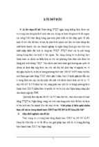
Figure 1. The MAPK signaling cascade. MAP3Ks are activated in response to stress,
cytokines or growth factors. Active MAP3Ks phosphorylate and activate MAP2Ks that in
turn phosphorylate and activate MAPKs. Active MAPKs activate cytosolic targets or
activate transcription factors that regulate the expression of genes that control different
cellular processes like proliferation, invasion, apoptosis and inflammation.
2
1.2 Characteristics and functions of MAPK proteins
MAPKs are proteins that are ubiquitously expressed in all eukaryotic cell types
but yet regulate different cellular responses in a stimulus- and cell type-specific manner
(Dhanasekaran and Johnson, 2007; Uhlik, et al., 2004; Widmann, et al., 1999). MAPKs
are proline directed serine/threonine kinases that activate nuclear or cytosolic substrates,
by phosphorylation at serine or threonine residues found within the Pro-X-Ser/Thr-Pro
consensus sequence (Alvarez, et al., 1991; Maeda and Firtel, 1997). MAPK proteins are
activated upon dual phosphorylation, by specific MAP2Ks, on both the threonine and
tyrosine residues of the Thr-X-Tyr motif present in the activation loop, where the amino
acid X varies with different MAPKs (Ahn, et al., 1991; D'Mello, et al., 1993; DiDonato,
et al., 1996; Estus, et al., 1994; Faris, et al., 1998; Frandsen and Schousboe, 1990).
MAPKs undergo an ordered phosphorylation mechanism, whereby the tyrosine residue of
the Thr-X-Tyr motif is phosphorylated first resulting in an increase in the affinity
between MAPKs and their specific MAP2Ks, a step that allows the subsequent
phosphorylation of the threonine residue and full MAPK activation (Haystead, et al.,
1992). Dual phosphorylation of MAPKs triggers a series of conformational changes in
the activation loop and surrounding sequences that ultimately result in the activation of
these proteins (Canagarajah, et al., 1997). Of the MAPK pathways, ERK, JNK and p38
signaling pathways are the best characterized.
1.2.1 The ERK1/2 pathway
ERK1 (44 kDa) and ERK2 (42 kDa), often referred to as ERK1/2, are two main
ubiquitously expressed isoforms of ERK that share more than 85% sequence identity
3
(Boulton, et al., 1991; Chen, et al., 2001; Seger and Krebs, 1995). Activation of ERK1/2
occurs upon specific recognition and subsequent phosphorylation of the Thr and Tyr
residues in the Thr-X-Tyr motif (Thr183 and Tyr185 in human ERK2 and Thr202 and
Tyr204 in human ERK1) by the upstream MAP2Ks, MEK1 and MEK2 (Crews, et al.,
1992; Zheng and Guan, 1993). Activity of ERK1/2 is also regulated by phosphatases,
including the MAPK phosphatases or MKPs which are dual-specificity phosphatases
(DSPs) that dephosphorylate both phospho-tyrosine and phospho-threonine residues
(Owens and Keyse, 2007; Raman, et al., 2007). Of the different MKPs, MKP3 shows
higher specificity towards ERK1/2 than other MAPKs (Zhang, et al., 2003). The
serine/threonine protein phosphatase 2A, or PP2A, also functions as a regulator of
ERK1/2 activity by dephosphorylating the threonine residue of the Thr-X-Tyr motif in
ERK1/2 (Anderson, et al., 1990).
Growth factors and mitogens are the primary activators of the ERK1/2 pathway,
however, cytokines, activators of G protein-coupled receptors and different stresses have
also been reported to activate this pathway (Johnson and Lapadat, 2002; Yoon and Seger,
2006). Early studies revealed a key role for Ras GTPases and B-Raf in ERK activation
(depicted in Figure 2 below). Briefly, receptor tyrosine kinases (RTKs), upon binding to
their ligands such as growth factors, undergo dimerization and cytoplasmic domain
transphosphorylation. Another transphosphorylation process then follows on specific
tyrosine residues in the cytoplasmic region of the RTK, which leads to full activation of
the receptor. The phospho-tyrosines create docking sites for the Src homology 2 (SH2)
domain of adaptor proteins, such as the growth factor receptor binding protein 2, or Grb2.
Grb2 then recruits the guanine nucleotide exchange factor (GEF), son of sevenless (SOS),
4
via its Src homology 3 (SH3) domain. SOS then activates Ras by promoting the switch
from an inactive GDP-bound to an active GTP-bound form (Buday and Downward,
2008; Downward, 1996; Wittinghofer, et al., 1997). Upon activation, Ras interacts with
and promotes the activation of members of the Raf family of MAP3Ks, Raf-1, B-Raf and
A-Raf. Once activated, Rafs phosphorylate and activate MEK1 and MEK2 that in turn
activate ERK1/2 (Chadee and Kyriakis, 2004; Dhillon, et al., 2007; Dunn, et al., 2005).
Active ERK1/2 will then undergo dimerization and either activate cytoplasmic substrates
such as p90 ribosomal S6 kinases (RSKs), mitogen and stress activated kinases (MSKs)
and MAPK interacting kinase (MNK), or translocate to the nucleus and regulate the
expression of certain genes by directly activating several transcription factors including
AP-1, c-Myc and c-Fos (Buday and Downward, 2008; Chen, et al., 2001; Dunn, et al.,
2005; Raman, et al., 2007).
5
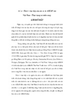
Figure 2. The Ras/Raf/ERK1/2 signaling pathway. In response to interaction with
growth factors (GF), RTKs undergo dimerization and activation. Grb2 binds to the active
RTK and recruits SOS which in turn activates Ras. Active Ras interacts with and
activates the Raf members. Active Rafs phosphorylate and activate MEK1/2 that in turn
phosphorylate and activate ERK1/2. Once activated, ERK1/2 can trigger a cellular
response either by activating cytoplasmic targets or by inducing transcriptional activation
by translocating to the nucleus and activating transcription factors.
6
- Xem thêm -